2.10.0
Hearing & Autism: a Polyvagal Misconception
In this chapter, we review polyvagal assumptions about hearing, reptiles, and autism. The PVT postulates that the reptilian ear (one bone instead of three) and the lack of “smart vagal” regulation (specifically cranial nerves V and VII) negatively affect the social life of “reptiles”. Autistic people often show reduced facial expression and social difficulties, which leads Porges to believe that their ventral vagal complex (VVC) — which includes the facial motor nerve CN V — is underactive. The next step is to put reptiles and autistic people side by side. This view is the theory behind the Listening Project (or Safe and Sound Protocol, SSD), which we will discover later (Auditory training).
2.10.1
Hearing and Auditory Reflexes
Hearing, or auditory, detects sound from vibratory motion transmitted through air or water. It is one of the traditional five senses, along with sight, touch, smell, and taste, and provides critical information about the environment necessary for survival. Parallel to the evolution of hearing (from a single ear bone in reptiles to three ear bones in mammals), various reflexes evolved to protect the inner ear from overstimulation by loud sounds (especially low-frequency). A continuous activity, as proposed by the PVT, is highly questionable.
Two Auditory Reflexes
The PVT postulates that the middle ear muscles (MEMR) selectively allow mammals to extract voice from noise. This should have allowed the first mammals to communicate at a higher sound frequency, unnoticed by reptilian dinosaurs. Porges is referring to a specific auditory reflex. What are these auditory reflexes?
The middle ear muscle reflex (MEMR) involves two tiny muscles in the middle ear: the stapedius muscle (CN VII) and the tensor tympani muscle (CN V). These muscles reduce the intensity of low-frequency sounds, preventing them from masking high-frequency sounds (such as the human voice). They also reduce the audibility of self-generated sounds such as speech, chewing, yawning, and sneezing.
The medial olivocochlear (MOC) reflex protects the basilar membrane (inner ear) from loud sounds. It involves cranial nerve VIII, responsible for both hearing and balance. MOC neurons can respond at much lower sound levels (Brown, 2009).
Because these reflexes protect hearing, especially from loud low-frequency sounds, some neuroscientists have long postulated (Borg, 1989) that they may prevent low-frequency sounds from masking higher-frequency sounds (e.g., speech). These hypotheses have not been confirmed. However, Borg's study is significant to Porges because it supports the PVT branchiomotor nerves CN V and CN VII, which are crucial for social life. He only recognized the first reflex for a long time, but recently, he admitted that CN VIII — the auditory part — could also be involved (Porges, 2014). Cortical involvement is still missing from the debate.
2.10.2
How does the Brain Extract the Voice from the Noise? Cortical Modulation of the Sounds
Scientific literature and reports
It is difficult to find precise answers to this question. However, below is a list of linked articles that may provide some interesting input:
How do our brains process speech? — Gareth Gaskell, a tutorial on YouTube
Pop-outs: How the brain extracts meaning from noise (Sanders, Robert, 2016).
Rapid tuning shifts in the human auditory cortex enhance speech intelligibility (Nature Communications)
Brain’s hippocampus helps fill in the blanks of language (Sept. 2016)
Brain’s iconic seat of speech goes silent when we actually talk (Feb. 2015)
Scientists decode brain waves to eavesdrop on what we hear (Jan. 2012)
2.10.3
Do Autistic People Hear Like Reptiles?
It is an odd question, but the Polyvagal Theory got us there. Reptiles have one ear bone instead of three (mammals). It has been said that their inability to filter out low-frequency sounds makes them asocial. From this point of view, autistic people have the same problem: they suffer from auditory hypersensitivity, their social life is poor, and their facial expression is limited. Do the math: autists have a reptilian issue! Don't they?
We will discuss auditory training and autism in more detail later. (Auditory Training). Porges published two articles about autism and hearing:
In Evolution and the Autonomic Nervous System: A Neurobiological Model of Socio-Emotional and Communication Disorders (Porges, Bazhenova, 2015, icdl.com/porges.html—no longer accessible), the authors describe an intervention using filtered music intended to trigger “the Social Engagement System.”
A new experiment was published under Reducing auditory hypersensitivities in autistic spectrum disorder: preliminary findings evaluating the listening project protocol in 2014 (Porges et al.)—more under auditory training.
2.10.4
Autism explained by Porges
Polyvagal Hypotheses
Autistic children have auditory hypersensitivity.
Reptiles have a smaller hearing range than mammals.
Reptiles have one middle ear bone (columella), while mammals have three.
Reptiles lack middle ear muscles to protect against loud sounds.
Middle ear muscles in mammals help extract individual voices from background noise, facilitating interspecies communication.
Reptiles and autistic children lack or have limited access to the audible range of frequencies corresponding to the human voice.
The polyvagal argument suggests that autistic children share similar auditory problems with reptiles.
A listening program using filtered music improves communication in autistic children.
After five consecutive 45-minute sessions, autistic children responded positively (e.g., opening their eyes and smiling).
This filtered music is believed to activate the vagal system and, consequently, the branchiomotor nerves V and VII.
As a result, HRV increases, which promotes sociability.
PVT provides both an explanation and a solution for autism.
Theoretical and Practical Critics
While some points may be valid — including the positive short-term outcome of this experience — many points are problematic.
Autistic children have a general hypersensitivity, e.g., tactile, visual, and auditory (low and high frequencies). This seems to be genetically caused, as researchers have been able to create strains of “autistic mice” through genetic manipulation.
The anthropocentric view is that a lower acoustic frequency is a problem. Every species has one mission: survival. Hearing ranges vary widely from species to species. Unlike birds, which rely on airborne sounds, many reptiles rely more on frequencies related to ground vibrations. Some have an excellent sense of smell (snakes).
Porges (2014) quotes Borg and Counter describing “the acoustic spectrum in our mechanized society (e.g., ventilation systems, traffic, airplanes, vacuum cleaners, and other appliances) resulting in both a hypersensitivity to sounds and distorting or “masking” the frequency components associated with human speech reaching the brain.” Considering that “modern” mammals are already 100 million years old, considerations based on 100 years of technology don't seem relevant. Except for the bellowing of dinosaurs and the occasional falling tree, the last million years must have been generally easy on the ears.
Some more limitations:
Most reptiles have middle ear muscles that attenuate loud sounds (Sánchez-Martínez, 2021).
No study has verified the role of the middle ear muscles in extracting the voice (of the mother). On the contrary, they protect the tympanum from loud, deep sounds. These sounds are not familiar in the natural environment except for sounds produced by the body. A damping of internal sounds has also been observed in some reptiles.
It's strange to propose that the reptilian auditory system is not adapted to its ecosystem, later implicitly suggesting that autistic children may have a similar problem as reptiles — to the point of hearing.
No robust study shows filtered music affects cranial nerves V and VII or HRV.
The specific setting (e.g., music + attention + quiet interaction and other factors described in the 2014 article) may induce a quiet response. The control period is not long enough. Other behavioral interventions were conducted in parallel with the filtered music sessions.
Limitations of the hypothesis: In Reducing Auditory Hypersensitivities (Porges, 2021, p. 226), Porges admits that “the hypothesized link between the middle ear transfer function and auditory hypersensitivity could be limited. Hypersensitivities, especially to high-frequency sounds, might be due not to the neural regulation of the middle ear muscles but to the olivary cochlear reflexes.”
2.10.5
Mammalian vs. Non-mammalian Hearing
According to the classical description, mammals have three middle ear bones and sauropsids (“reptiles” and birds) have one. Functionally, this is incorrect, as many reptiles (lizards) and birds have a bipartite middle ear (see below).
The delicate bone structure of the mammalian ear did not appear from the beginning. Instead, it appeared at least twice, as evidenced by its appearance in a species that lived during the dinosaur era some 123 million years ago. This repeated appearance underscores the topic's evolutionary importance and complexity.
Further Reading
In Comparative Anatomy of the Middle Ear in Some Lizard Species with Comments on the Evolutionary Changes within Squamata (2021), Sánchez-Martínez describes the middle ear of various “lizards.” Like most reptiles, they have two structures: the columella — a tiny bone with a foot inserted into the oval foramen — and the extracolumella — a cartilaginous structure inserted into the tympanum (eardrum). A hinge connects the two structures. The extracolumellar muscle, attached to the cartilaginous part, regulates the intensity of vibrations and protects the hearing system from loud sounds. Some species have an external muscle for physical and sound protection. Birds have the same organization. Crocodilians also have a muscle attached directly to the eardrum. Birds have a muscle (M. Tensor tympani) that pulls on the tympanic membrane and the extracolumella.
In owls with exceptional hearing, examination of this muscle shows reflex contraction to loud sounds. See Peripheral Hearing Mechanisms in Reptiles and Birds (Manley, G.A., 1990, p. 38).
Major Evolutionary Transitions and Innovations: The Tympanic Middle Ear (Tucker, 2017).
The Middle Ear of Reptiles and Birds (Saunders, 2000)
Size does matter: Crocodile mothers react more to the voice of smaller offspring (Chabert, 2015).
Automated acoustic identification of individuals across species: improving identification across recording conditions (Stowell, 2019). Individual differences in acoustic signals have been widely reported in all classes of vertebrates (e.g., fish, amphibians, birds, and mammals). This article discusses how different species have evolved their discriminative systems, including the acoustic context (e.g., river sounds).
Middle ear muscles of the frog (Wever, 1979) describes how a frog’s middle ear muscles enable it to control sound reception, protecting the inner ear from overstimulation caused by its calls and the loud chorus of neighboring frogs.
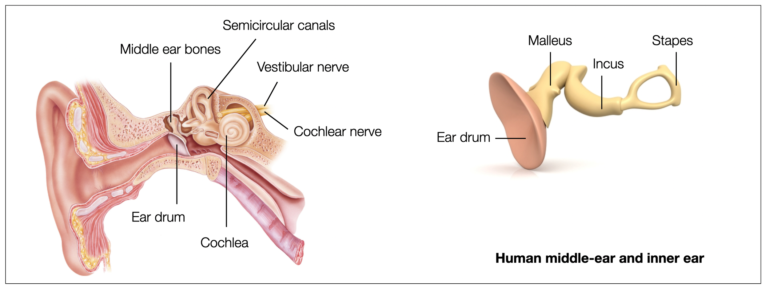
Auditory System: Efferent Systems to the Auditory Periphery (Brown, 2009) shows how MOC (medial olivocochlear) neurons and stapedius motor neurons respond to sound. MOC neurons can respond to much lower sound levels than the stapedius (75-90 dB). OC neurons receive input from higher centers (including the auditory cortex). Flexibility within the vertebrate middle ear (Mason, 2013) provides a detailed comparative description of different vertebrate middle ear models (see diagrams below). The authors argue that “the chain of conducting elements is flexible: even the ‘single ossicle’ ears of most non-mammalian tetrapods are functionally ‘double ossicle’ ears due to mobile articulations between the stapes and extrastapes (…) The most obvious role of middle-ear flexibility is to protect the inner ear from high-amplitude displacements.”
The first diagram shows a human middle ear — three bones. The second diagram shows the elaborate middle ear of the lizard. The second element (extracolumella) is cartilaginous. Birds have a similar organization.
2.10.6
Autism (ASD) and Autonomic Nervous System
In The vagus: A mediator of behavioral and physiologic features associated with Autism (Porges, 2005, p.72), Porges writes, “the literature suggests that autism is associated with reliable differences in the amplitude of respiratory sinus arrhythmia and the transitory heart rate response pattern to various stimuli and task demands (…) These findings, describing compromised cardiac vagal regulation in autistic individuals, support predictions from the Polyvagal Theory. Consistent with the Polyvagal Theory, both visceromotor (i.e., vagal) and somatomotor (e.g., eye gaze and facial expression) components of the Social Engagement System are compromised in individuals with disorders such as autism.
An aberrant precision account of autism (Lawson, 2014) gives an interesting description of the way autistic people process information, with difficulties in properly encoding information (e.g. visual), leading to sensory overload and difficulties in contextualizing their perceptions.
Neurobiology of social behavior abnormalities in autism and Williams syndrome (Barak, 2016)
Autism Spectrum Disorder in Children Is Not Associated With Abnormal Autonomic Nervous System Function: Hypothesis and Theory (Barbier, 2022) presents a critical review of the polyvagal view, with which she strongly disagrees.
Autonomic Dysfunction in Autism Spectrum Disorder (Owens, 2021) describes multiple autonomic problems in a small population with ASD. However, dysregulation plays a role in both ways (hyper- and hyposympathetic tone). “In ASD, basal heart rate and responses to orthostatic tests of autonomic function were elevated, supporting previous findings of increased sympathoexcitation. However, sympathetic vasoconstriction was impaired in ASD. Conclusion: Intermittent neuro-cardiovascular autonomic dysfunction affecting heart rate and blood pressure was over-represented in ASD.“
Autonomic Responses to Head-Up Tilt Test in Children with Autism Spectrum Disorders (Bricout, 2018) concludes: “Our results showed that children with ASD did not have clinical signs of dysautonomia in response to a head-up tilt test. However, in children with ASD higher parasympathetic responses with the same sympathetic modulations during orthostatic stress sug- gest parasympathetic dominance in this population (…) our results suggested that children with ASD do not have clinical autonomic dysfunction per se in response to the head-up tilt test, because no clinical sign of dysautonomia was observed either at rest or during this test.”
2.10.7
Autism and Genetics
PVT reduces autism spectrum disorders to a lack of auditory regulation caused by a lack of middle ear muscle activity. Cranial nerves V and VII, parts of the ventral vagal complex, are involved. Porges never considers genetic causes, which have been clearly identified in the scientific literature in humans and animal models. (Wilde, 2022, Csillag, 2022). (Barak, 2016; Takarae, 2017; Roussel, 2022),
Idiosyncratic connectivity in autism: developmental and anatomical considerations (Uddin, 2015) describes particular patterns of connectivity by autistic persons, leading to an impaired filtering of sensory inputs.
Precision Autism: Genomic Stratification of Disorders Making Up the Broad Spectrum May Demystify its “Epidemic Rates” by Torres (2021). Illustration.
SFARI.org and DisGeNET.org are Genetic Databases. SFARI is a division of the Simons Foundation. SFARI ‘a mission is to improve the understanding, diagnosis, and treatment of autism spectrum disorders by funding innovative research of the highest quality and relevance.
Peripheral Somatosensory Neuron Dysfunction: Emerging Roles in Autism Spectrum Disorders (Orefice, 2020).
2.10.8
Further Reading on Autism and Hearing
Auditory and opioids
According to Panksepp (2012, p. 306), the auditory system is incredibly rich in opioids (...) Fetuses begin to integrate extra-uterine sounds and even recognize their mother's voice before birth.
Butler and Hodos (2005, p. 214) describe the acoustic reflex as follows: “Two muscles are attached to the ossicles: the stapedius (facial VII) and the tensor tympani (V, trigeminal). These muscles and the ossicles help to dampen intense sounds, thereby preventing damage to the acoustic receptors in the inner ear.”
Cortical contributions to the perception of loudness and hyperacusis (McGill, 2023): “Sound perception is closely linked to the spatiotemporal patterning of neural activity in the auditory cortex (ACtx). Inhibitory interneurons sculpt the patterns of excitatory ACtx pyramidal neuron activity and thus play a central role in sculpting the perception of sound. Reduced inhibition through inhibitory interneurons and increased gain of sound-evoked pyramidal neuron spike rates are consequences of aging and sensorineural hearing loss” (quote from abstract).
Heart rate variability in individuals with autism spectrum disorders: A meta-analysis (Cheng, 2020) “The results support low HRV to be a potential biomarker of ASD, especially RSA reactivity under social stress” (quote from abstract).
Domestic cat larynges can produce purring frequencies without neural input (Herbst, 2023)
Highest sound: Bats 20 kHz (ultrasound), Golden-winged Manakin 11.9 kHz, highest piccolo (instrument) 4.6 kHz,
Lowest birdsong 0.2 kHz, 0.04 lowest double bass, purring cat 0.02 kHz
In this article, which focuses on purring cats, the author challenges the view that mammalian vocalizations are systematically located in the mid to high frequencies.
Hyperacusis in Autism Spectrum Disorders (Danesh, 2021) “Utilizing Magnetic Resonance Imaging (MRI), scientists have seen elevated auditory activity in the auditory midbrain, thalamus and cortex, as well as enlarged subcortical and cortical responses to sound in subjects with hyperacusis. (…) Auditory integration training has little credible data demonstrating its effectiveness. Increased awareness of hyperacusis, expansion of the body of literature, and improvements with research design over time have led to stronger evidence-based intervention approaches for this condition, and ongoing research is warranted to continue improving best practices” (quote from abstract).
>> to the next chapter Dissociation


