2.0
Vocabulary of the Neuroanatomy
In the following sections, we will give you some basic information about the nervous system, you may need for better understanding the PVT.
Neuroanatomy is the most complicated part of the anatomy because, during evolution, the initial simple neural tube folded many times and expanded in all directions. Maps on the paper — and the visual system, anyway — are two-dimensional. But the brain is three-dimensional. Like all painters, scientific illustrators must use shadows, perspective, and colors to create a suggestive representation of the neural system. But, even hard-working medical students have trouble learning the structures of the brain.
2.1.1
The Brain
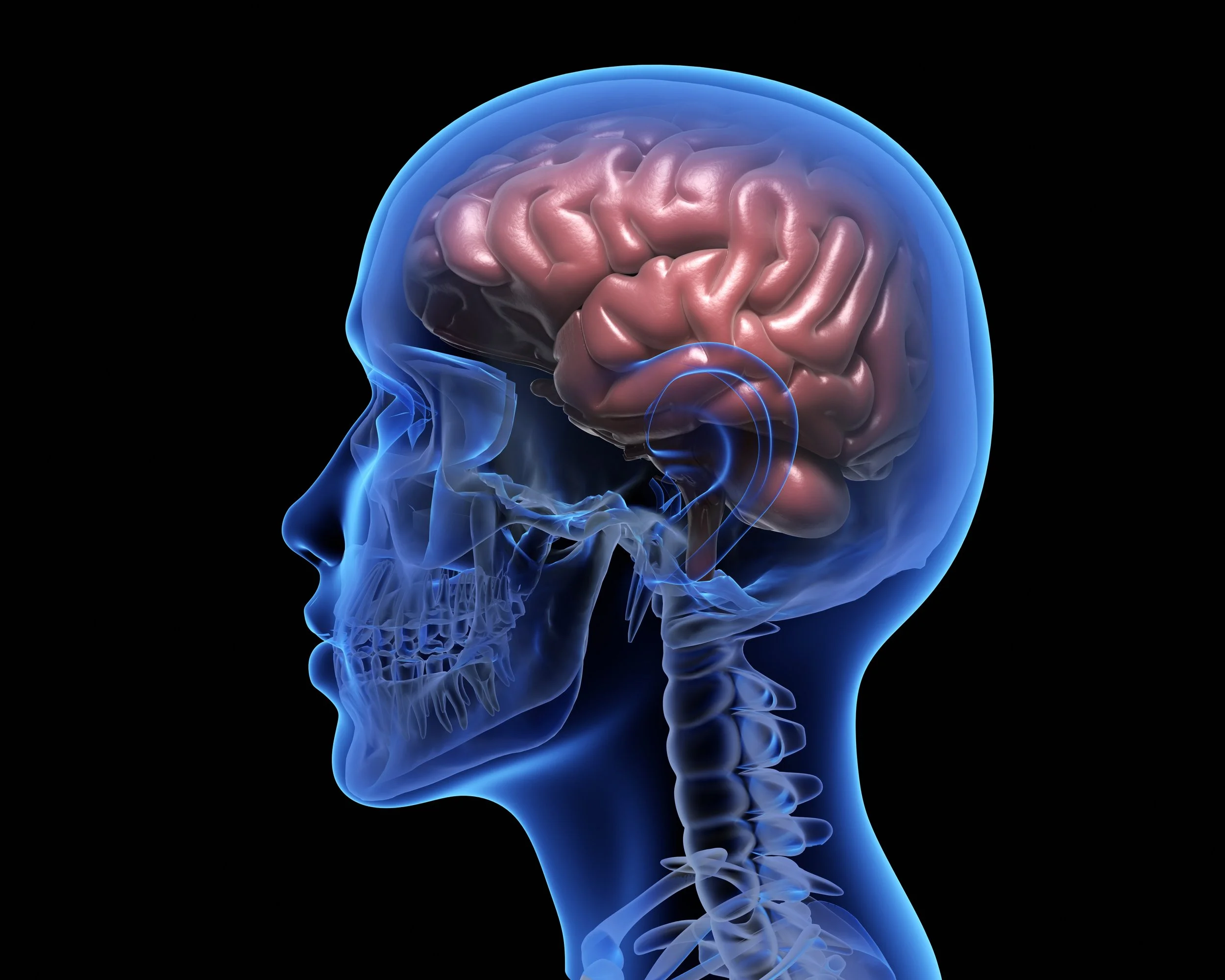
The brain contains 86.1 billion neurons and 84.6 billion glial cells.
Get the directions!
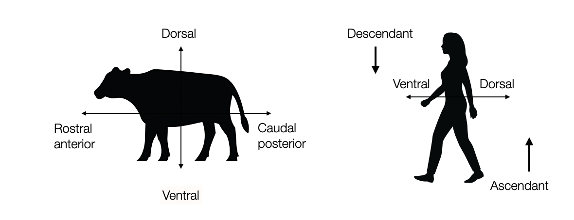
Humans walk upright — unlike most animals. The inferior-superior axis (animal) becomes anterior-posterior. The brain stem is half vertical. To avoid confusion, anatomists call the anterior part rostral (Latin: rostrum = beak of a war galley) and the posterior part caudal (Latin: cauda = tail). They also use the terms ventral (belly, abdomen) and dorsal (back) — hence ventral vs. dorsal vagal.
2.1.2
Neurons
Simple cells communicate. Plants communicate, too. They send biochemical signals. But about 600 mya, the animal world sees the appearance of a new form of cell – the neurons – allowing an electric propagation of the signal over extremely long distances – like the invention of the telegraph in the nineteenth century.
Neurons have a nucleus and a cell body. What makes them unique are the short (dendrites) and long (axon) extensions, which serve to communicate with other neurons or with organs. The nervous system is the ensemble of all neurons.
2.1.3
Cortex, nuclei, and ganglion
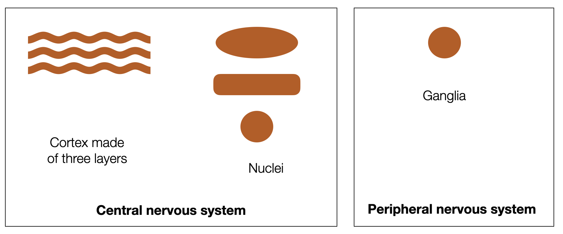
In the brain, bodies of neurons are grouped into:
layers (cortex)
nuclei, which are mostly round, oval, or elongated formations (columns).
At the periphery, outside the brain, neuron bodies form ganglions (or ganglia)
2.1.4
Nervous System
The nervous system have three general functions:
Sensory (afferent, ascending)
Motor (efferent, descending)
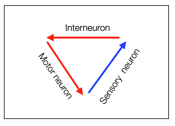
Integrative (interneurons)
It’s divided into
the Central Nervous System, protected by a bony structure (skull or vertebrae)
the Peripheral Nervous System
2.1.5
Nerve Fibers
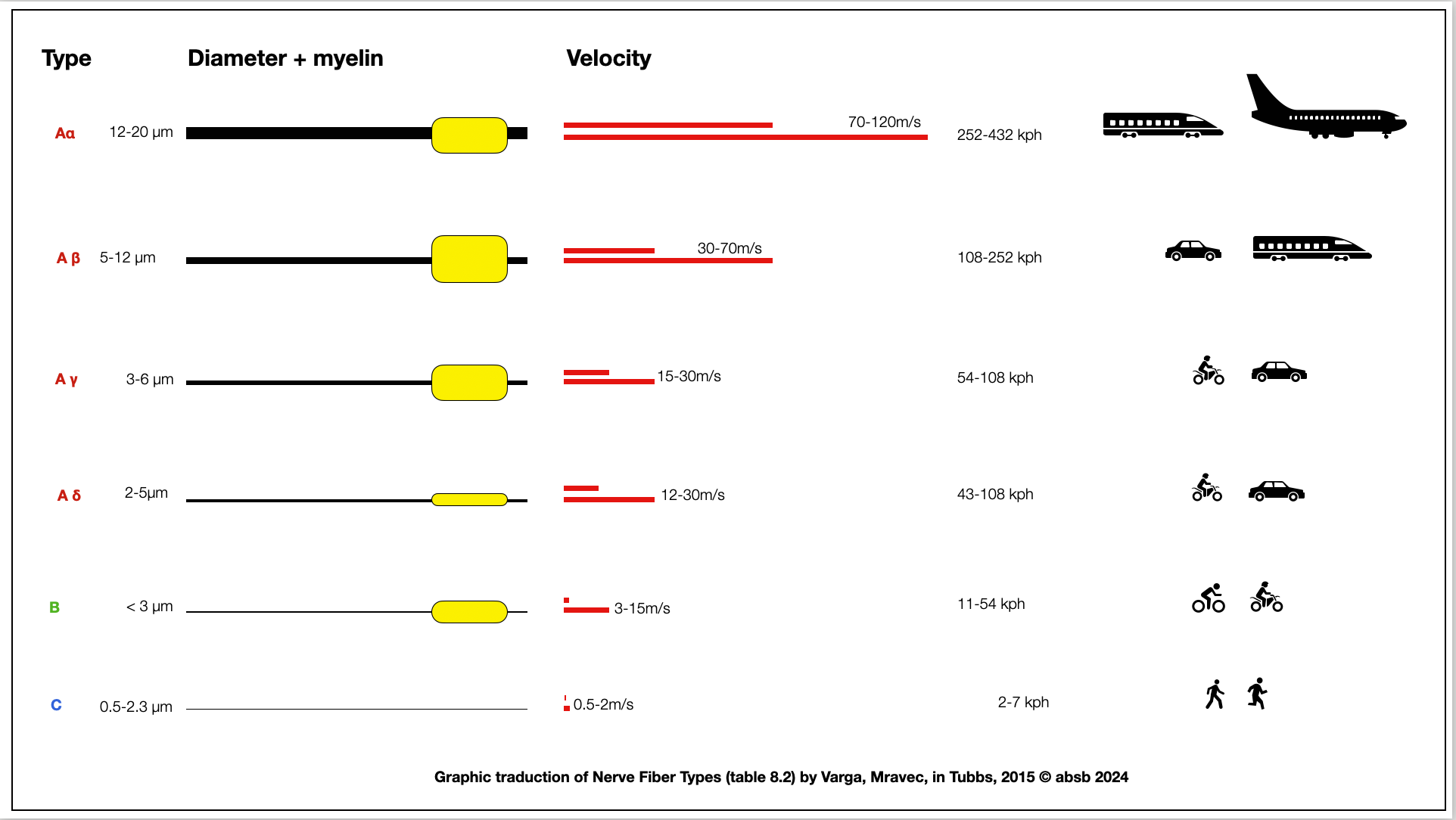
Nerve fibers generally refer to a single axon, including its sheath. They vary in diameter and speed. Some are myelinated, and some are not. To illustrate their velocity, we compared the speed of signal transmission to that of a person walking, a car, or an airplane.
Based on Nerve Fiber Types (Varga, Mravec in Nerves and Nerves Injuries (Tubbs, 2015).
-
The terms "axons," "fibers," and "nerves" refer to different aspects of the nervous system.
Axons: An axon is a long, slender projection of a neuron, that typically conducts electrical impulses away from the neuron's cell body. Axons can vary greatly in length, from a tiny fraction of an inch to several feet in humans (as in the case of axons running from the spinal cord to the toes).
Fibers: The term "fiber" is often interchangeable with "axon" but can also refer to a bundle of axons.
However, “fiber” generally refers to the axon + the sheath formed by the glial cells (with or without myelin).
Nerves: A nerve is a bundle of many axons, frequently thousands, wrapped together in a protective sheath. Nerves are the main components of the peripheral nervous system (PNS), which extends outside the central nervous system (CNS) to connect it with limbs and organs. Nerves can be sensory, motor, or mixed (carrying both sensory and motor fibers).
-
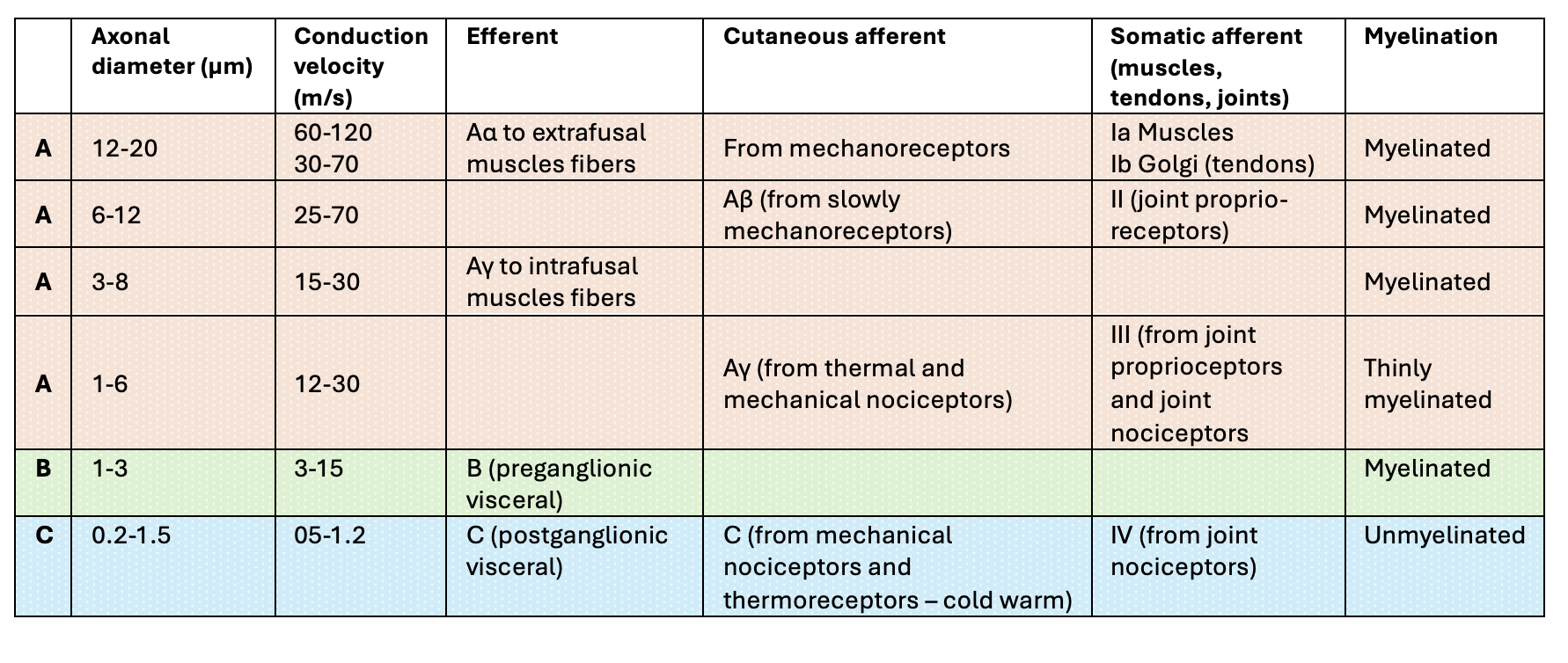
A group is subdivided into four types (A-alpha, A-beta, A-delta, and A-gamma fibers) based on the information carried by the fibers and the tissues they innervate.
A-alpha fibers are the primary receptors of the muscle spindle and Golgi tendon organ.
A-beta fibers act as secondary receptors of the muscle spindle and contribute to cutaneous mechanoreceptors.
A-gamma fibers are typically motor neurons that control the intrinsic activation of the muscle spindle.
A-delta fibers are free nerve endings that conduct painful stimuli related to pressure and temperature. The myelinated A-delta fibers respond to mechanical (pressure) stimulus and produce the sensation of sharp, localized, fast pain.
-
Fibers of the B group are myelinated with a small diameter and have a low conduction velocity. The primary role of B fibers is to transmit the parasympathetic (motor, efferent) information from the ventral part of the Nucleus Ambiguus to viscera (heart, lungs). They are also found in the dorsal parasympathetic branch (DMVN).
-
Fibers of the C group are unmyelinated, have a small diameter, and low conduction velocity. The lack of myelination in the C group is the primary cause of their slow conduction velocity. The unmyelinated C fibers respond to thermal, mechanical, and chemical stimuli and produce the sensation of dull, diffuse, aching, burning, and delayed pain.
Peripheral nerves, based on the excellent website NYSORA.
2.1.6
Myelinated nerves
Speed is of the essence for the nervous system — as for any messaging service! The evolution of myelinated nerves, a significant milestone in the vertebrate lineage, revolutionized the nervous system. From the first jawless vertebrates to fish, the emergence of myelinated nerves has played a crucial role in survival strategies, whether as prey or predator. This evolution of the myelination program in vertebrates, as studied by Zalc (2008), is a fascinating journey to explore.
Myelinated nerves, with their unique ability to increase speed, are a marvel of the nervous system. While it's true that myelinated fibers are not always energy efficient and take up more space, their role in rapid signal transmission is unparalleled. The fact that not all fibers express this property, such as the sensory C-fibers responsible for pain and pleasure, adds a fascinating layer to the nervous system's complexity.
To use the road metaphor again, let's compare myelination to road tar. According to the Federal Highway Administration (FHWA), as of 2019, the United States has approximately 4.4 million miles of public roads. Approximately 2.7 million miles were paved, while the remaining 1.7 million miles were unpaved or surfaced with materials other than asphalt or concrete. So, do we need myelin? Not always.
When and why did myelin appear in evolution? Myelin, the fatty substance that insulates nerve fibers and improves the speed of electrical impulses along neurons, is not a recent innovation. It first appeared in evolution about 500 million years ago during the Cambrian explosion, a period that marked the rapid diversification of multicellular life. This early appearance of myelin, coinciding with the first complex nervous system, is evidence of its critical role in the evolution of life.
Myelin is a characteristic feature of vertebrates (animals with a backbone, such as fish, amphibians, reptiles, birds, and mammals) and some invertebrates, including certain annelids (segmented worms) and crustaceans. The presence of myelin in these diverse groups suggests that it appeared early in the evolution of Bilaterians (bilateral symmetry and central nervous system).
What determines whether a nerve is myelinated or not? What role do glial cells play? Are some nerves partially myelinated?
Several factors determine whether a nerve is myelinated, including its specific function, location, and the signals it needs to transmit. Myelinated nerves are more efficient at conducting electrical signals quickly over long distances, which is essential for coordinating movement, processing sensory information, and regulating complex physiological processes.
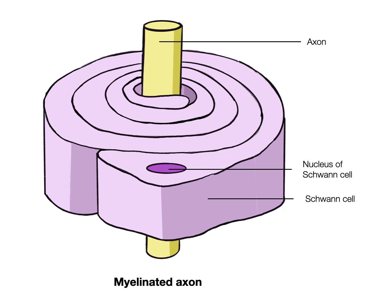
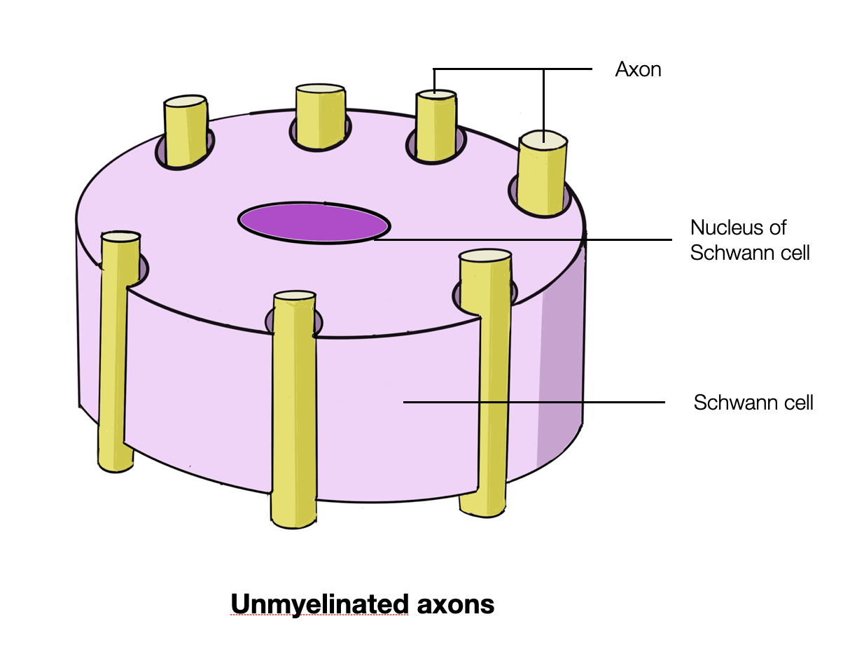
Unmyelinated axons are not “naked” but protected and bundled by Schwan's cells. A fiber is an axon plus the protecting sheet (myelinated or unmyelinated. A group of fibers forms a nerve.
Glial cells, non-neuronal cells in the nervous system, play a critical role in the formation and maintenance of myelin. In the central nervous system (CNS), including the brain and spinal cord, oligodendrocytes are the glial cells that produce myelin. In the peripheral nervous system (PNS), which includes nerves outside the CNS, Schwann cells are responsible for myelination. These glial cells wrap around the axons of neurons, forming layers of myelin that insulate the nerve fibers and facilitate the rapid transmission of electrical signals.
Myelin is interrupted at regular intervals by unmyelinated segments called nodes of Ranvier. The nodes of Ranvier are critical for the fast and efficient conduction of electrical signals because they allow the action potential (the electrical impulse) to jump from one node to the next, a process called saltatory conduction. This results in faster signal transmission than an unmyelinated nerve fiber.
Several functions of the nervous system do not require fast signaling, such as regulating internal organs or perceiving certain types of pain. Unmyelinated fibers have a slower conduction speed than myelinated fibers but can transmit signals effectively for their specific functions. More on Nysora.
2.1.7
Somatic vs. Autonomic Nerve Fibers
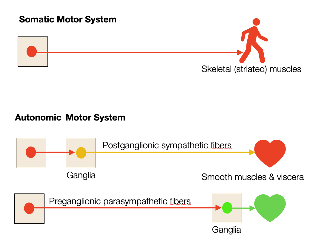
In the scientific description of the autonomic system, you often come across the terms “preganglionic vs. postganglionic.” What does this mean? Somatic motor fibers (innervating striated muscles) go directly to the muscles. While their bodies are inside the CNS (brain or spinal cord), their long axons are outside. Therefore, they are part of the peripheral nervous system. They don't have a stop in the ganglia.
On the contrary, all autonomic fibers (sympathetic and parasympathetic) have a two-part organization. The preganglionic neuronal bodies are inside the CNS, and their axons are outside. These fibers connect (synapse) with postganglionic neurons in the ganglia. The bodies of postganglionic neurons form masses called ganglia.
All somatic motor fibers and preganglionic fibers are cholinergic, and the neurotransmitter is acetylcholine. The sympathetic postganglionic nerves are adrenergic, and the neurotransmitter is noradrenaline (norepinephrine).
There are several ways to block the nervous system. Nerve agents such as sarin block the destruction of acetylcholine, leaving the receptors in a state of continuous activation. Soldiers use an injection of atropine to block the acetylcholine receptors. Atropine is also a general antimuscarinic blocker (M1-M5). It blocks the vagal (parasympathetic) stimulation of the heart. Propranolol, a “beta-blocker,” blocks the sympathetic stimulation—it is the drug of choice for stressed performers on stage.
2.1.8
Nerves
A nerve fiber is the long, slender projection of a nerve cell, or neuron, that typically conducts electrical impulses away from the neuron's cell body. The term “fiber” can refer to a single axon or dendrite, whereas a “nerve” is a larger structure containing many axons or nerve fibers, often bundled with connective tissue and blood vessels.
The main differences between a nerve and a nerve fiber are
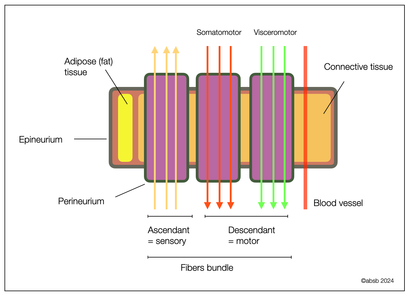
Composition: A nerve is a bundle of many nerve fibers, blood vessels, adipose, and connective tissue, whereas a nerve fiber is a single axon or dendrite of a neuron (with its protective sheet).
Function: Nerves are like highways, allowing traffic in both directions (motor = descendant vs. ascendant = sensory) on parallel lanes (fibers).
Structure: Nerves are larger structures that can be seen without a microscope, while fibers are microscopic and consist of individual axons. Nerves can be seen and manipulated during surgery or osteopathic treatment. Nerve fibers are microscopic components.
Illustration of the anatomy of a typical peripheral nerve. Available from: https://www.researchgate.net/figure/llustration-of-the-anatomy-of-a-typical-peripheral-nerve_fig1_330757070
2.1.9
Afferent vs. Efferent Nerves
The peripheral nervous system is divided into afferent (sensory) and efferent (motor) nerves. The afferent or sensory division transmits impulses from peripheral organs to the CNS. Their bodies are grouped into ganglia outside the CNS. The efferent or motor nerves transmit impulses from the CNS to the peripheral organs (e.g., muscles). In humans, sensory pathways are ascending, and motor pathways are descending.
This diagram shows afferent nerves (green) and efferent motor nerves (purple). We usually find interneurons between the incoming sensory and outgoing motor pathways.
2.1.10
Cranial vs. Spinal Nerves
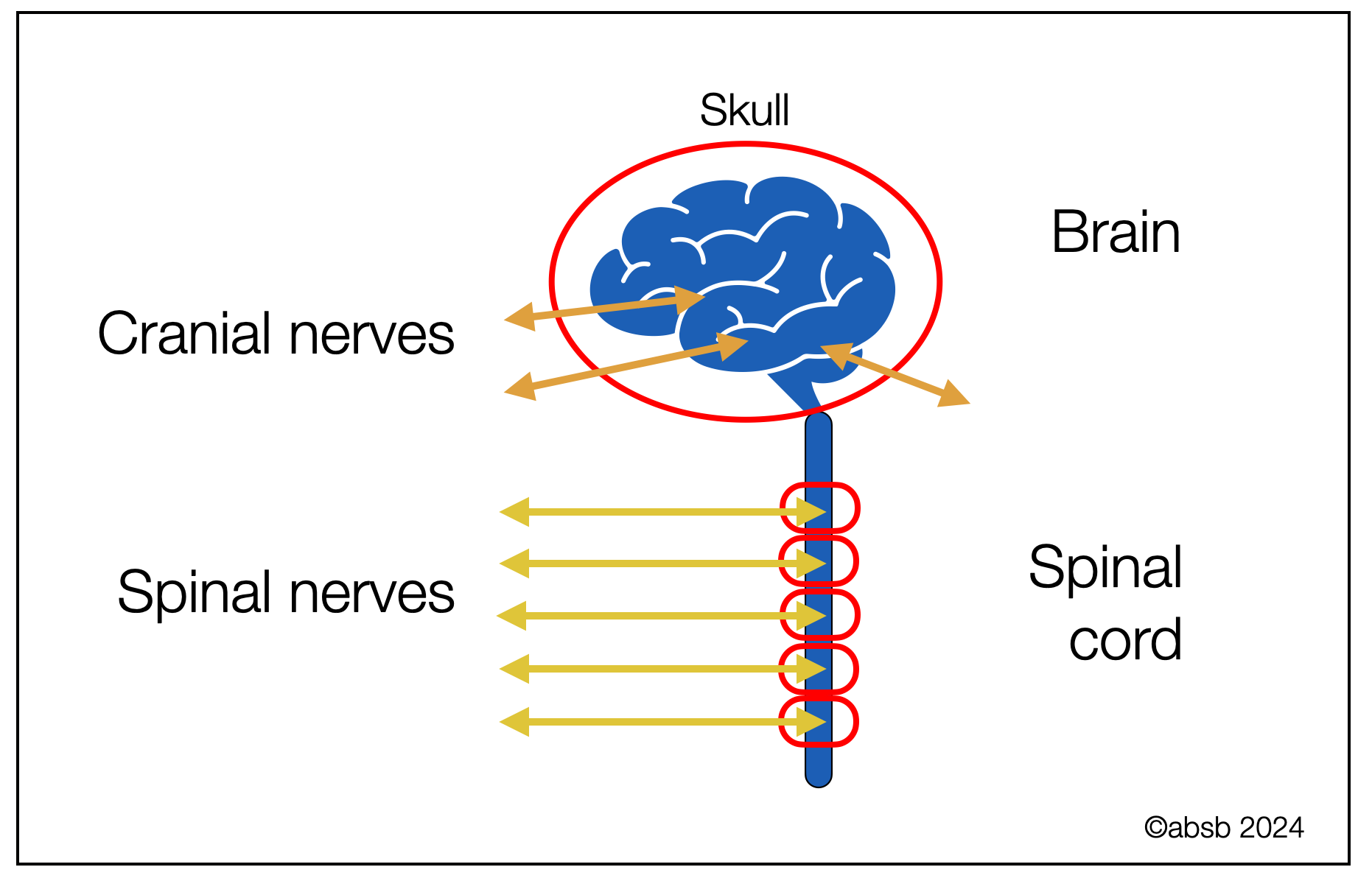
The twelve paired cranial nerves exit the CNS through the cranium (skull). They generally innervate organs in the head (e.g., nose, eyes, ears, or throat). The CN X (vagus nerve) is an exception, as it also innervates the viscera (e.g., lungs, heart, or intestines).
Spinal nerves leave the CNS at the level of the spine. They refer to striated muscles (e.g., biceps) or sensory information (e.g., proprioception and skin).
2.1.11
The Central Nervous System
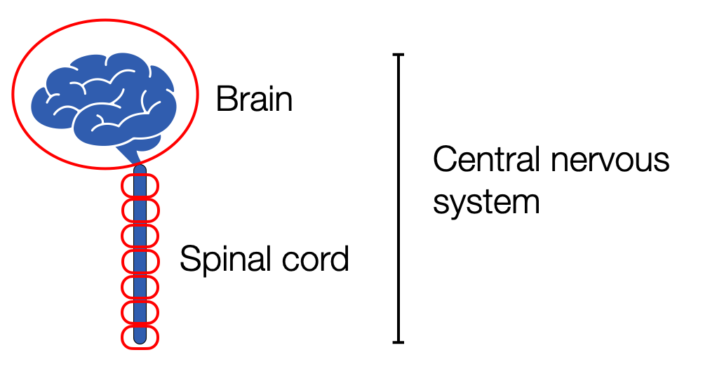
The brain and spinal cord are the organs of the central nervous system. Because they are so important, they are protected by bones: the skull and the spine. The brain and spinal cord share a continuous fluid-filled cavity: the brain's ventricles (or chambers) and the spinal canal.
2.1.12
The Peripheral Nervous System
The organs of the peripheral nervous system are nerves and ganglia. Nerves are bundles of nerve fibers. It is important to remember the difference between nerves and fibers. Just as a highway may have several lanes with different speeds and directions, a single nerve may contain:
afferent (ascending) sensory and/or efferent (descending to muscles or viscera) motor fibers
A-B-C and/or fibers (different diameters and speeds)
myelinated and/or unmyelinated fibers
somatic (related to striated muscles and skin) and visceral (related to organs and smooth muscles)
The vagus nerve is the perfect example of a mixed nerve because it contains all of these variations.
Fibers are not nerves; they are part of them. They are like the many lanes of a large highway. Some lanes are slower, some are faster. Some go up, some go down. Similarly, the vagus nerve has 80% upstream (sensory) and 20% downstream (motor). The motor ratio is highly variable: fast branchiomotor fibers to the striated muscles (swallowing and phonation), slow parasympathetic fibers to the heart and lungs, and very slow fibers to the digestive system. There are even some sympathetic fibers. So we should think of a crossroads rather than a long road.
A nerve such as the vagus nerve can contain up to six types of fibers: motor (to striated muscles), visceral motor (sympathetic, parasympathetic B and C), visceral afferent, and sensory afferent!
2.1.13
Somatic vs. Visceral Systems
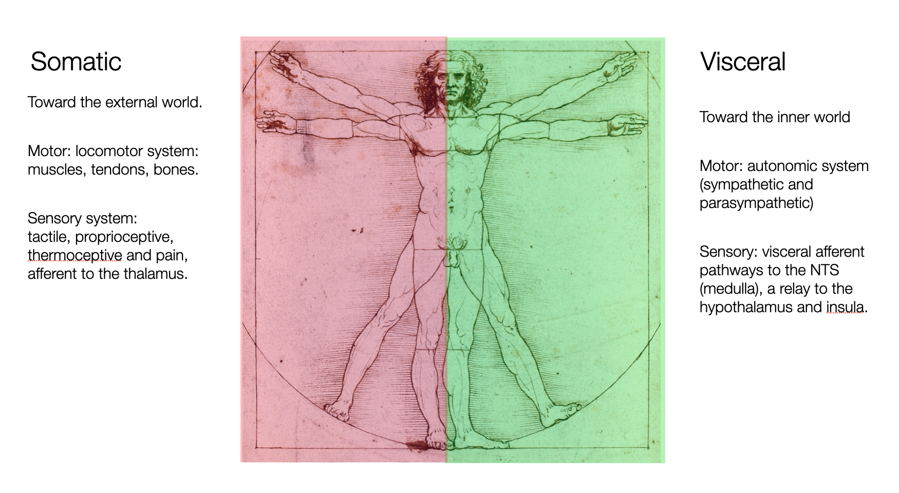
The Somatic Nervous System (SNS) innervates the voluntary muscles (e.g., skeletal or striated muscles).
The Visceral Nervous System (VNS) comprises the efferent motor Autonomic Nervous System (ANS) and the General Afferent Visceral nervous system.
The main difference between the two efferent systems is how their fibers synapse to their target organ:
Somatic efferent fibers have a direct route from the CNS (brain or spinal cord) to the nicotinic receptors on muscles.
Visceral efferent fibers have a two-step route: preganglionic visceral efferent fibers activate nicotinic receptors in the ganglia (relays) and synapse on postganglionic fibers, which activate:
Sympathetic: adrenergic receptors (noradrenaline).
Parasympathetic: muscarinic receptors (acetylcholine).
BW background = Man of Vitruve by da Vinci, by Paris Orlando, colors and texts by absb.
The somatic nervous system (SNS) controls the body's voluntary movements. It consists of 43 segments (Rea, 2014), each of which consists of a motor (efferent) nerve to command muscle contraction and a sensory nerve to relay information to the central nervous system (CNS). The motor and sensory parts form a reflex arch.
12 cranial nerves pass directly into or out of the skull
31 spinal nerves (cervical, thoracic, lumbar, sacral, and coccygeal) enter and exit the spinal cord.
At different levels (cranial or spinal), autonomic efferent nerves (sympathetic or parasympathetic) and visceral afferent nerves may travel with somatic nerves (mixed nerves).
The somatic nervous system and the five senses give us a place in the world. Information gathered from receptors in muscles, tendons, and joints makes us aware of our body every second. However, a significant amount of movement is automatically managed outside of awareness. In addition, muscle tone (tension) can be difficult to control. Apart from substances (e.g., drugs, medicine, or alcohol), the only way to relax is through massage, meditation, hypnosis, or yoga. This takes time.
The visceral nervous system gives us access to the inner world of our internal organs: the heart, lungs, and digestive system. The motor part, or autonomic nervous system (ANS) (Langley, 1921) describes the sympathetic, parasympathetic, and enteric nervous systems. , The general visceral afferent fibers (GVA), which make up about 80% of the vagus, conduct sensory information from the viscera to the Nucleus Tractus Solitarius (NTS) are neither sympathetic nor parasympathetic, but the sensor part of the visceral nervous system.
Polyvagal confusion
The somatic SNS and the autonomic ANS are separate systems. Porges confuses them when he identifies the dorsal vagal complex as “the source of the most primitive defensive behavior”: “immobilization — freezing or involuntary playing dead, with loss of consciousness and muscle tone — to minimize detection” (Porges, 2021, p. 205). The dorsal vagal complex DVC, consisting of three groups of neurons (DVMN, NTS, and AP), is involved in visceral actions. None of them understands “freezing, playing dead, or with loss of muscle tone” as these behaviors are all related to the somatic system, commanding the skeletal muscles.
2.1.14
The Visceral Nervous System
The visceral nervous system connects the CNS (brain stem or spine) to the peripheral organs or viscera (e.g., lungs, heart, digestive system). It is also involved in regulating the immune system.
We can distinguish between the motor part (= autonomic nervous system), which consists of the sympathetic, parasympathetic, and enteric nervous systems, and the visceral afferent system.
A. Autonomic Nervous System (ANS)
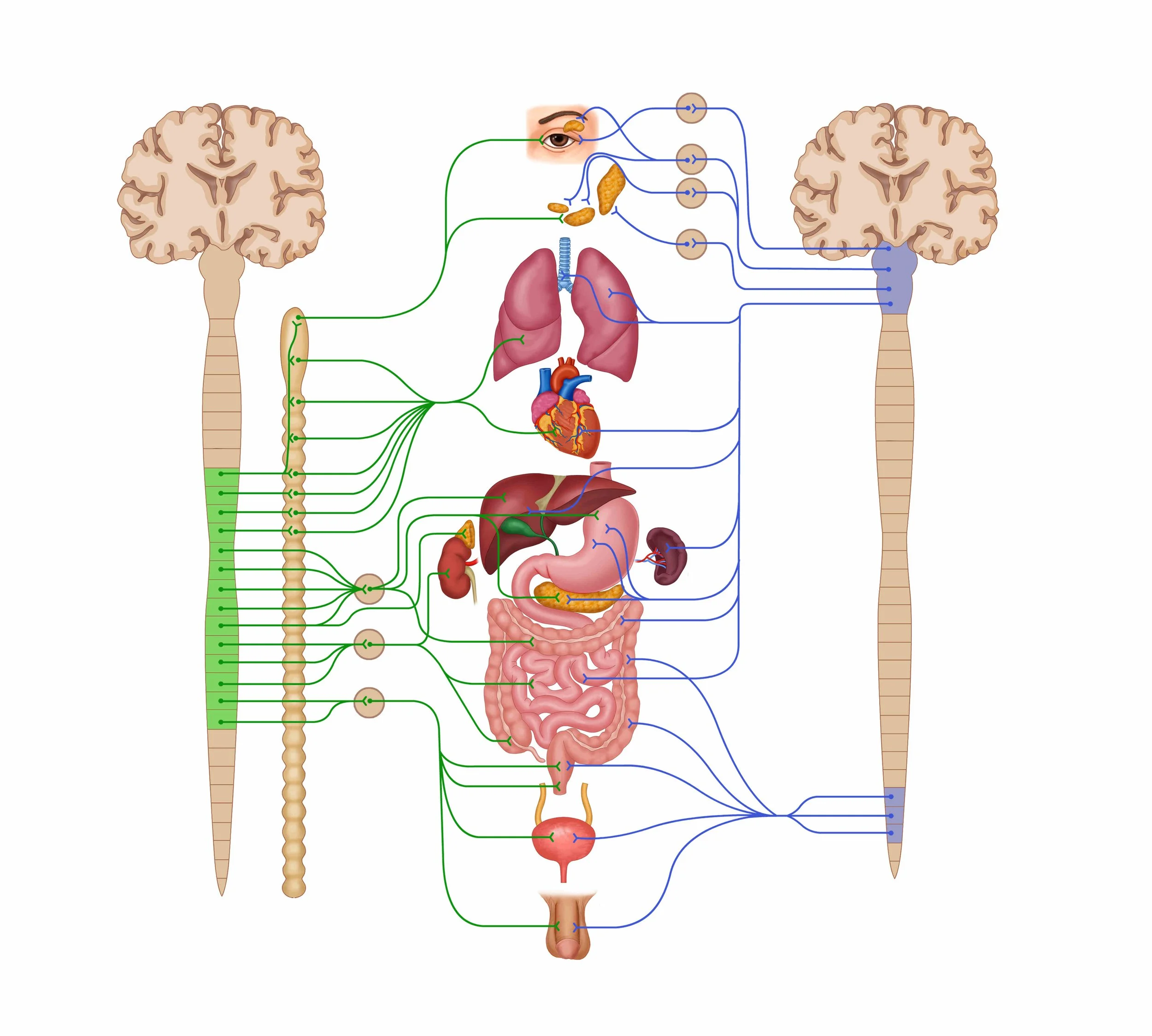
The ANS has three divisions, each with a specific anatomical organization and physiological function. Unlike the spinal nerves, these nerves have an additional relay outside the spine at the level of the ganglia. There, preganglionic nerves synapse with postganglionic nerves.
1. Sympathetic Nervous System SNS
The sympathetic ganglia form a double vertical chain of ganglia proximal to the thoracic and lumbar spine. The postganglionic nerves are adrenergic (noradrenaline or norepinephrine).
2. Parasympathetic Nervous System PNS
The parasympathetic ganglia are located proximal to the target organs.
In the upper part of the system, preganglionic nerves exit at the level of the brainstem (parasympathetic portions of cranial nerves III, VII, IX, and X). The tenth nerve extends to the heart, lungs, and digestive system.
In the second part, the preganglionic nerves exit at the level of the sacrum and innervate the bladder, sexual organs, and the last part of the colon.
Postganglionic nerves are cholinergic (acetylcholine, muscarinic receptors).
3. Enteric Nervous System (END)
As the third division of the autonomic nervous system, the enteric nervous system (Greek: enteron = intestine) represents a self-organized digestive system. It has little to do with the CNS. Some writers call it “the second brain,” which is incorrect, while its computational capacity — compared to the “first brain” — is minimal.
B. Visceral Afferent System
Made up of slow C-fibers (slow) and faster A-delta fibers, it collects information about the internal organs (lungs, heart, digestive system). The ascending system brings information to the Nucleus of Tract Solitary (NTS) in the brain stem, which is processed and sent to other centers.
In green: sympathetic system,
in blue: parasympathetic system.
2.1.15
Cranial Nerves
The cranial nerves (CN) are a set of twelve paired nerves that originate or terminate within the cranium (skull) (Butler, 2005, Chap. X-XII). The nerves are:
purely sensory (I, olfactory, II ophthalmic, and VIII vestibular—auditory).
purely motor (III, IV, VI oculomotor)(XI and XII)
or mixed (V, VII, IX, and X)
Finally, the cranial nerves III, V, IX, and X have a parasympathetic component.
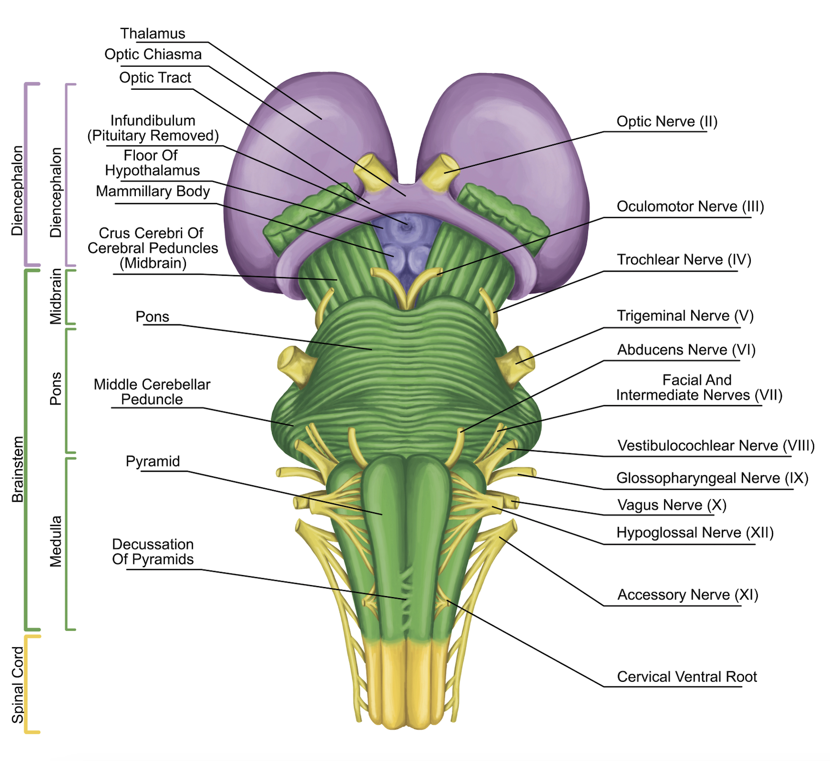
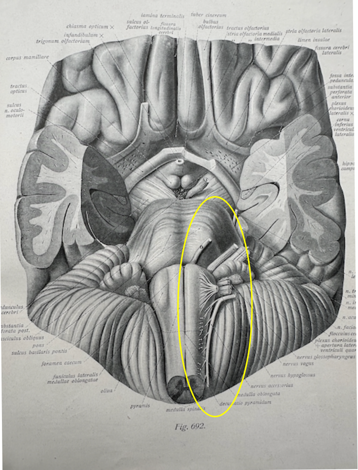
Here we see a part of the cranial nerves leaving the brain from the anterior (inferior) part of the brainstem (yellow oval). (Original figure from Sobotta, 1922).
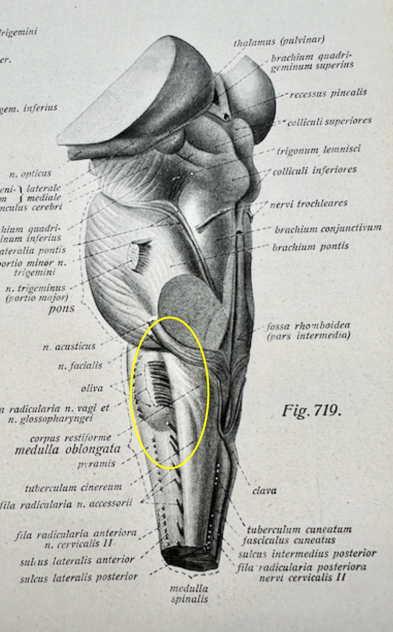
Nerve roots of the IX, X, and XI (from the Nucleus Ambiguus) on a lateral view of the brain stem. (Original figure from Sobotta, 1922).
2.1.16
Vagal Nerve
The tenth cranial nerve (X) is the longest and most complex of the cranial nerves. The vagal nerve is called because of its wandering appearance (Latin: vagus = wanderer). It is a mixed nerve:
80% afferent, ascending (sensory) fibers that carry visceral sensory information (e.g., heart, lungs, intestines, liver, pancreas) to the brainstem (NTS)
20% efferent, descending (motor) fibers originate from neurons in the dorsal motor nucleus of the vagus and nucleus ambiguus.
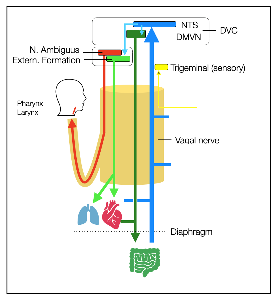
This diagram shows a more systematic view of the vagus nerve. We want to make the reader aware of the ambiguity of the word “vagal.
In many texts, “vagal” refers to the parasympathetic fibers — dorsal and ventral — whose terminals synapse (connect) with cholinergic muscarinic receptors (e.g., heart or intestines).
“Vagal tone” describes activity in the vagal nerve (yellow area), but it's unclear how and where this activity is measured. Some scientists propose to equate HRV (which only reflects the input of the NAext) with vagal tone. Grossman and Taylor (2007) disagree.
Classically, the vagal nerve is thought to arise from four nuclei (Baker, 2022): Nucleus ambiguus, NTS, DVMN, and trigeminal. This description does not include the peripheral ganglia (Nodosum and Jugular), which contain the cell bodies of the afferent vagal fibers. On the other hand, neither the NTS nor the AP (which, together with the DVMN, form the so-called Dorsal Vagal Complex or DVC) is “vagal”: their territory doesn't extend beyond the central nervous system. The NTS is the primary target of peripheral afferent fibers that synapse via glutamatergic or GABAergic receptors on second-order neurons of the NTS.
“Parasympathetic” is often confused with “vagal,” although parasympathetic fibers make up less than 20% of the vagal nerve. The parasympathetic system also includes some fibers of cranial nerves III, VII, and IX and the sacral parasympathetic fibers (bladder, genitals, and last part of the intestines).
The vagal nerve contains somatomotor (to striated muscles of the larynx and pharynx) and somatosensory (ear) fibers, which are not parasympathetic.
The sensory auricular nerve, which terminates in the trigeminal nucleus (cranial nerve V), has no visceral function, although it briefly shares its path with visceral fibers.
2.1.17
Medulla
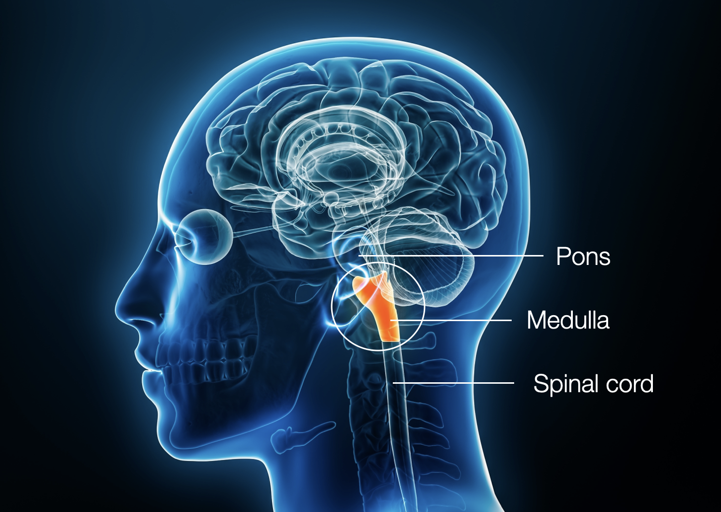
The medulla oblongata (myelencephalon) is the lowest part of the brain stem. It is home to the “ventral” and “dorsal” sources of the vagal nerve. The other centers of the VVC (Ventral Vagal Complex) are located in the pons (=bridge), just one level higher: CN V (trigeminal) and CN VII (facial) (branchiomotor nerves). The ambiguus nucleus has two components: a branchiomotor part that innervates the pharynx (swallowing) and larynx (vocalization) and a general visceral part that innervates the heart and lungs.
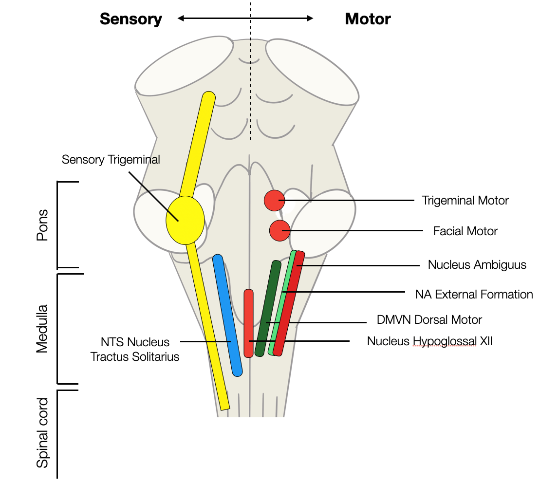
Most of the structures we will discuss are located in the medulla, the lowest part of the brain. In the figure, on the left, you can see the sensory (afferent) nuclei (trigeminal = CN V and NTS). They bring (afferent) information from the face (V) and the viscera (NTS), respectively. The nuclei on the left are motor: in red, three branchiomotor nuclei innervating the jaws (V), the face (VII), and the pharynx-larynx (IX-X). The hypoglossal somatomotor nucleus innervates the tongue. The (ventral) external formation of the NA and the (dorsal) DVMN are the sources of the two parasympathetic branches.
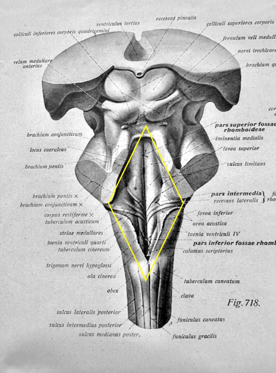
(Original figure from Sobotta, 1922). Notice the rhomboid yellow shape.
(Original figure from Sobotta, 1922). Notice the triangular yellow shape describing the fourth ventricle.
The medulla forms the rhombencephalon with the pons, which develops around the fourth ventricle. This chamber has a pyramidal structure, combining a rhomboid and a triangular shape (as shown in yellow in the following figures). The orange rectangle at midbrain level marks the location of the PAG (periaqueductal gray matter). Anatomists must section the cerebellum to access the fovea oblongata (fourth ventricle).
2.1.18
Parasympathetic Ventral vs. Dorsal
The Parasympathetic System The word para is a Greek suffix with a double meaning:
Along or beside, as in parallel, paramilitary, paravertebral, paralympic, or paramedical.
Opposite, as in parachute, paradox (against the principle), or paranoia (against healthy thinking).
Accordingly, the sympathetic and parasympathetic systems work in parallel or in opposition, depending on the situation, to ensure the maintenance of the "internal environment" (Bernard, 1878) or homeostasis (Cannon, 1929).
VCV and DVC
The neologism “ventral vagal complex"(VVC) describes a group consisting of the nuclei of the branchiomotor (pharyngeomotor) cranial nerves CN V, VII, IX, X, and XI. This term is used only by polyvagal authors. CN XI (accessory) is a special case because it joined the group phylogenetically later. Moreover, only a tiny part is cranial (medullary). The most important part has its roots in the spinal cord (spinal accessory), but it shares genetic characteristics with the branchiomotor nerves (Ryan, 2007), (Benninger, 2010) and (Tada, 2015).
The DVC (Dorsal Vagal Complex), a more recent term, includes three nuclei: the NTS, the DMVN, and the AP. Some authors use this term to describe interactive circuits (e.g., satiety), including a sensory ascending vagal branch (synapsing at the NTS) and a motor descending vagal branch (the DMVN). The area postrema (AP) interacts with the NTS for metabolic regulation.
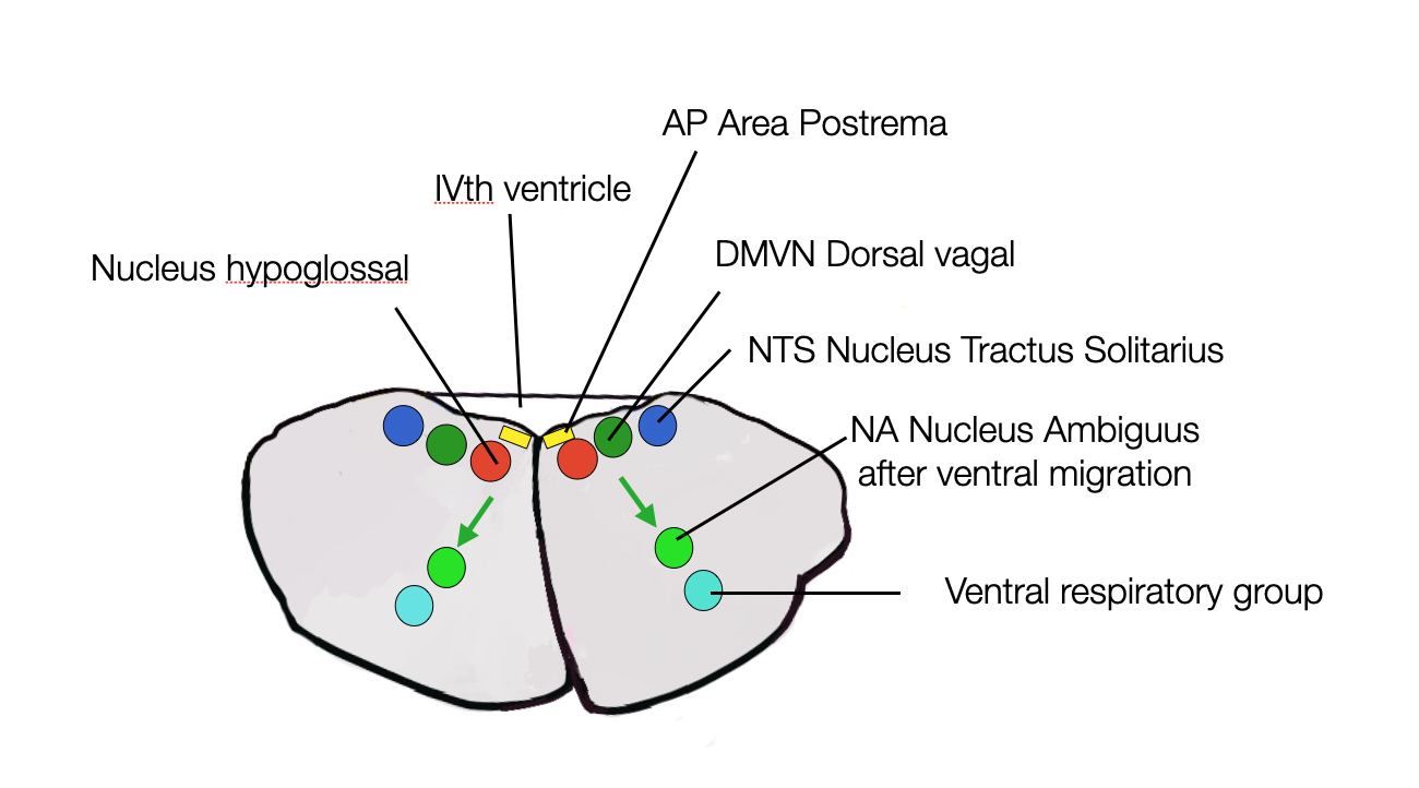
This section of the brain stem (at the level of the medulla) shows the ventral and dorsal regions.
2.1.19
Orthovagal vs. Polyvagal – the new stamp
Our research aims to describe the autonomic nervous system (ANS) according to the most recent knowledge (2024). Since our findings in the scientific literature were constantly contradicting the “polyvagal” postulates, we searched for a term for “scientifically current.” Since the sympathetic system is sometimes called “orthosympathetic,” we chose “orthovagal.” The ancient Greek word ορθοσ = orthos means correct or upright, as in orthography, orthopedic, or orthostatic. This website will use the term “orthovagal” to emphasize a particular description that fits the current literature but not the PVT. We will develop our arguments about this in later chapters.
>> to the next chapter Ontogeny & Phylogeny
